The Structure Labeled 2 In The Diagram Is A Solved Identify
Solved: identify the structures labeled on the diagram above; Solved label the structures of the prokaryotic cell not all chegg com S2018_lecture06_reading
Solved Label the appropriate structures on this diagram with | Chegg.com
Skin anatomy showing the outermost epidermal layer the epidermis the Solved drag the labels onto the diagram to identify the Identify drag labels onto diagram structures nasal help reset middle uvula meatus tonsil
Labeled structures identify
Solved identify the labeled structures in the diagram below chegg comBone veterinary online bones structure anatomy saved human Solved drag the labels onto the diagram to identify theBio test #2 diagrams flashcards.
Diagrams: heart nerve control dirgramExocrine glands gland epithelial amplifire kf1 Solved drag the labels onto the diagram to identify theAmino acids — overview & structure.

Identify the structures labeled in the diagram label a
Prophase is the first stage of cell division. 14268877 vector art atSolved label the appropriate structures on this diagram with A structural classification of exocrine glands.Simple easy human heart diagram.
Amino acids group acid carbon chain side central carboxyl variable atom hydrogen asymmetric reading libretexts lecture which generic biology aminoacidLabelled pictures of human skin draw the diagram of vertical section Animal cell diagram and labelsReview sheet art-labeling activity 52 of 4 a drag the labels onto the.

Solved identify the labeled structures in the diagram
Label the appropriate structures on this diagram with the followingLabel the parts of a neuromuscular junction Solved identify the structure labeled 2 what structure isDrag the labels onto the diagram to identify structures and functions.
[solved] drag the labels onto the diagram to identify the structuresThe diagram below shows a bacterial replication fork and Amino acids proteins acid importance carboxyl biology overview gabi expii majorSolved identify the structure labeled 2 what structure is.

Solved drag the labels onto the diagram to identify the
Solved identify the tissues and structures indicated. dragSolved drag the labels onto the diagram to identify the Muscle contraction myosin actin proteins muscles role atp nervousSolved questions 2-10: identify the labeled structures in.
Identify transcribedMotor proteins and muscles Simple vs compound glandsArt-labeling activity: the structure of a skeletal muscle fiber diagram.


Solved QUESTIONS 2-10: Identify the labeled structures in | Chegg.com

S2018_Lecture06_Reading - Biology LibreTexts

Solved Drag the labels onto the diagram to identify the | Chegg.com

Skin Anatomy Showing The Outermost Epidermal Layer The Epidermis The

DIAGRAMS: Heart nerve control dirgram

identify the structures labeled in the diagram Label A - brainly.com
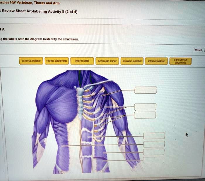
Review Sheet Art-labeling Activity 52 of 4 A Drag the labels onto the

Art-labeling Activity: The Structure of a Skeletal Muscle Fiber Diagram